The Prostate Protocol By Scott Davis The Prostate Protocol is designed for all those who want a natural solution for BPH. The online program can help users to treat BPH. Also, it will address the root cause and prevent a recurrence. You might not expect this benefit from conventional treatments. The program is the outcome of extensive research. You can download this program and use it for a lifetime. There will be no additional costs. Also, you do not need to spend on other things to support your health. Moreover, you will have the money refund option.
Structure of the prostate gland
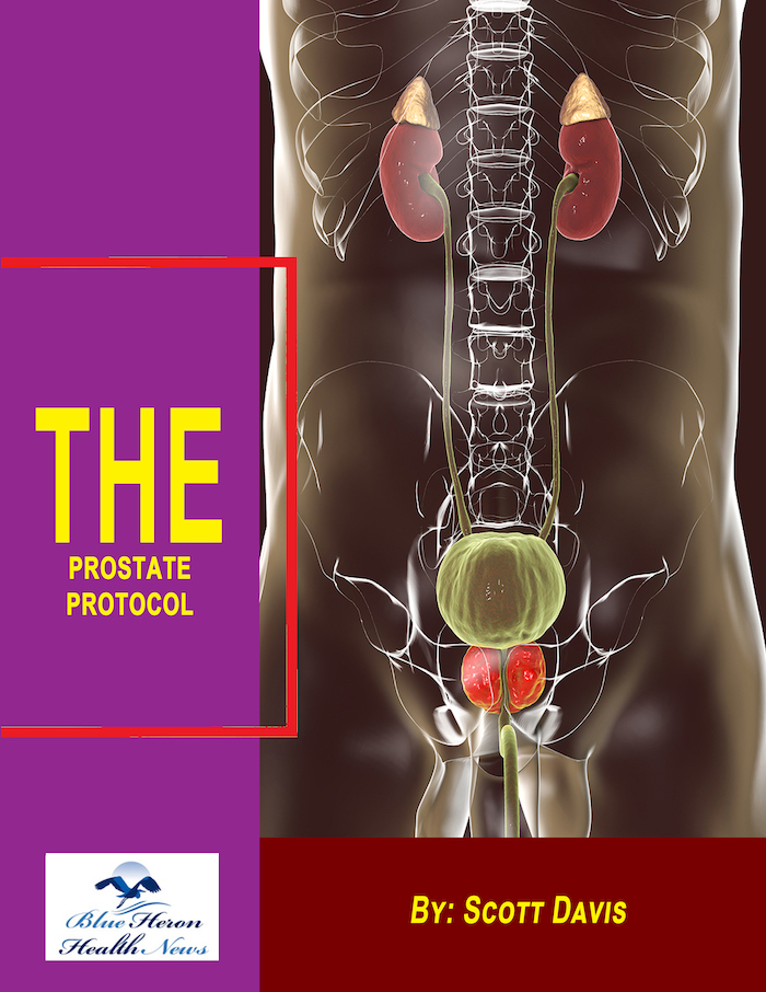
The prostate gland is a small, walnut-sized organ located in the male pelvis, just below the bladder and in front of the rectum. It surrounds the urethra, the tube through which urine and semen pass out of the body. The structure of the prostate gland can be understood by examining its anatomy, zonal structure, and histology.
1. Anatomy
- Size and Shape: The prostate is approximately the size of a walnut, with an average weight of 20-30 grams in a healthy adult male. It is conical in shape, with its base located at the bladder neck and its apex pointing downward toward the urogenital diaphragm.
- Location: The prostate is situated just below the bladder and encircles the prostatic urethra, which is the portion of the urethra that runs through the prostate.
2. Zonal Structure
- Peripheral Zone (PZ): This is the largest zone, comprising about 70% of the prostate gland. It is located at the back of the prostate and is the area most commonly affected by prostate cancer.
- Central Zone (CZ): This zone surrounds the ejaculatory ducts and comprises about 25% of the prostate. It is less commonly involved in prostate cancer but is more resistant to it when compared to the peripheral zone.
- Transitional Zone (TZ): This is the area around the urethra and accounts for about 5% of the prostate volume in young men. It is the zone where benign prostatic hyperplasia (BPH) most commonly occurs, leading to urinary symptoms.
- Anterior Fibromuscular Stroma: This area consists mainly of muscle and fibrous tissue and is located at the front of the prostate. It does not contain glandular tissue.
3. Histology
- Glandular Tissue: The prostate gland is made up of numerous small glands that produce prostatic fluid, a component of semen. These glands are lined with a layer of epithelial cells and are surrounded by smooth muscle fibers that help expel the fluid during ejaculation.
- Stroma: The stroma of the prostate consists of connective tissue, smooth muscle fibers, and blood vessels. The smooth muscle fibers contract during ejaculation to push the prostatic fluid into the urethra.
- Capsule: The prostate is enclosed in a thin, fibrous capsule that helps maintain its shape and provides some protection. The capsule also contains blood vessels and nerves.
4. Vascular Supply
- Arterial Supply: The prostate receives blood supply primarily from the inferior vesical arteries, which branch from the internal iliac arteries. Additional blood supply comes from the middle rectal and internal pudendal arteries.
- Venous Drainage: Blood is drained from the prostate via the prostatic venous plexus, which surrounds the prostate and connects to the internal iliac veins.
5. Lymphatic Drainage
- Lymph from the prostate drains into the obturator and internal iliac lymph nodes, which are key sites for the spread of prostate cancer.
6. Innervation
- The prostate is innervated by the autonomic nervous system, specifically the sympathetic and parasympathetic nerves. Sympathetic nerves are responsible for the contraction of smooth muscle during ejaculation, while parasympathetic nerves influence glandular secretion.
The prostate’s structure plays a crucial role in its function, particularly in the production of seminal fluid and in the control of urinary flow. Its complex anatomy and proximity to critical structures like the bladder and urethra are also why prostate diseases can have significant impacts on both urinary and reproductive health.
Certainly! Here’s a more detailed exploration of the structure of the prostate gland:
1. Anatomy of the Prostate Gland
- Size and Shape:
- The prostate is a chestnut-shaped organ, approximately 3 cm long, 4 cm wide, and 2 cm thick. It weighs about 20-30 grams in an adult male, though its size can vary depending on age and the presence of conditions like benign prostatic hyperplasia (BPH).
- It is divided into two lobes (right and left) that are connected by an isthmus (or median lobe), which lies in front of the urethra.
- Location:
- Base: The base of the prostate is the superior portion, which is adjacent to the bladder neck. The base is wide and flat.
- Apex: The apex is the narrow, inferior portion of the prostate, which rests on the urogenital diaphragm and is directed downward toward the perineum.
- Posterior Surface: This part of the prostate lies against the rectum, making it accessible for digital rectal examination (DRE). It is the area where many prostate tumors are palpated.
- Anterior Surface: This surface is adjacent to the pubic symphysis and is covered by the puboprostatic ligaments that anchor the prostate to the pelvic floor.
- Relations:
- Superiorly: The prostate is continuous with the base of the bladder.
- Inferiorly: It is connected to the external urethral sphincter and perineal muscles.
- Anteriorly: The pubic symphysis and retropubic space (space of Retzius) lie anterior to the prostate.
- Posteriorly: The prostate is related to the rectum, separated by the rectovesical fascia (Denonvilliers’ fascia).
- Laterally: It is flanked by the levator ani muscles, part of the pelvic floor.
2. Zonal Structure of the Prostate
The prostate gland is divided into distinct zones, each with different physiological and pathological characteristics:
- Peripheral Zone (PZ):
- Constitutes about 70% of the prostate’s volume.
- Located posteriorly and laterally, surrounding the distal urethra.
- Contains the majority of the glandular tissue, making it the primary site for prostate cancer. About 70-80% of prostate cancers originate here.
- The peripheral zone can be palpated through a digital rectal examination (DRE).
- Central Zone (CZ):
- Comprises about 25% of the prostate’s volume.
- Surrounds the ejaculatory ducts and is positioned posteriorly near the bladder base.
- Less commonly involved in cancers but is more resistant to inflammation and malignancy compared to the peripheral zone.
- Has a distinct embryological origin, which may contribute to its resistance to cancer.
- Transitional Zone (TZ):
- Accounts for about 5% of the prostate volume in young men but can enlarge significantly in older men due to benign prostatic hyperplasia (BPH).
- Surrounds the urethra just below the bladder and is the area most commonly affected by BPH, leading to urinary symptoms such as frequency, urgency, and weak stream.
- BPH nodules primarily develop in this zone, causing compression of the urethra and resulting in obstructive urinary symptoms.
- Anterior Fibromuscular Stroma:
- Located at the anterior aspect of the prostate, this zone contains no glandular tissue, consisting mainly of dense fibromuscular tissue (smooth muscle and fibrous tissue).
- This zone is relatively resistant to pathological changes like cancer or BPH.
3. Histology of the Prostate
The prostate is histologically composed of both glandular and stromal elements:
- Glandular Tissue:
- The glandular component consists of numerous tubuloalveolar glands, organized into lobules, which are responsible for producing prostatic fluid, a key component of semen.
- The glandular epithelium is typically simple columnar but can vary from cuboidal to pseudostratified, depending on the functional state.
- Secretory Acini: These are the terminal portions of the glandular ducts that secrete the prostatic fluid. The acini are surrounded by a basal layer of low cuboidal or flattened cells and a luminal layer of columnar secretory cells.
- Ductal System: The glands drain into ducts that converge and open into the prostatic urethra. The ducts are lined by a similar epithelium to the acini and are supported by smooth muscle and connective tissue.
- Stroma:
- The stromal component is a mixture of fibrous connective tissue and smooth muscle fibers.
- Smooth muscle contraction during ejaculation aids in expelling prostatic fluid into the urethra.
- The stroma also contains blood vessels, lymphatic vessels, and nerve fibers, contributing to the gland’s complex vascular and neural regulation.
- Capsule:
- The entire prostate gland is encased in a dense, fibrous capsule, which extends septa into the gland to create lobules.
- This capsule is not a true anatomic capsule but rather a condensation of the pelvic fascia that also covers the seminal vesicles and bladder.
4. Vascular Supply
- Arterial Supply:
- The prostate receives blood primarily from the inferior vesical arteries, which are branches of the internal iliac arteries.
- Additional blood supply may come from branches of the middle rectal and internal pudendal arteries.
- The arterial supply is crucial for maintaining the gland’s function and is also a consideration during surgeries such as prostatectomy.
- Venous Drainage:
- The venous blood is drained through the prostatic venous plexus, which surrounds the prostate.
- This plexus connects with the deep dorsal vein of the penis and the internal iliac veins.
- The prostatic venous plexus can be a route for cancer metastasis to other organs, particularly the vertebral column.
5. Lymphatic Drainage
- Lymphatic drainage from the prostate is primarily directed to the internal iliac lymph nodes.
- Additional drainage occurs to the external iliac, obturator, and sacral lymph nodes.
- The lymphatic system is important in the spread of prostate cancer, and involvement of these nodes can indicate advanced disease.
6. Innervation
- The prostate is innervated by both the sympathetic and parasympathetic divisions of the autonomic nervous system.
- Sympathetic Innervation: Derived from the hypogastric plexus, it is responsible for the contraction of the smooth muscles during ejaculation, leading to the release of prostatic fluid.
- Parasympathetic Innervation: Provided by the pelvic splanchnic nerves (S2-S4), which modulate the glandular secretion of the prostate.
- Sensory Innervation: The prostate also receives sensory nerve fibers that can transmit pain signals, which are relevant in conditions like prostatitis.
7. Function of the Prostate
- Semen Production: The prostate contributes 20-30% of the seminal fluid, which includes enzymes, prostate-specific antigen (PSA), citric acid, zinc, and other components that support sperm viability and motility.
- Regulation of Urinary Flow: The prostate’s location around the urethra allows it to play a role in the regulation of urine flow, especially under the influence of hormonal changes or pathological conditions like BPH.
- Enzyme Secretion: The prostate secretes proteolytic enzymes, including PSA, which liquefy semen after ejaculation, allowing sperm to swim freely.
This detailed understanding of the prostate gland’s structure is crucial for comprehending how various diseases, such as BPH, prostatitis, and prostate cancer, develop and affect the gland’s function.

The Prostate Protocol By Scott Davis The Prostate Protocol is designed for all those who want a natural solution for BPH. The online program can help users to treat BPH. Also, it will address the root cause and prevent a recurrence. You might not expect this benefit from conventional treatments. The program is the outcome of extensive research. You can download this program and use it for a lifetime. There will be no additional costs. Also, you do not need to spend on other things to support your health. Moreover, you will have the money refund option.